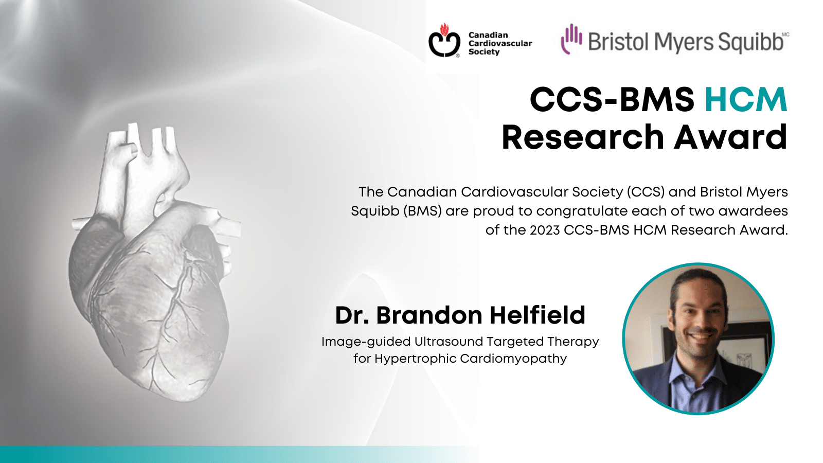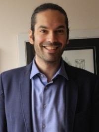

Dr. Brandon Helfield, an Assistant Professor at Concordia University in Montreal, is the winner of a CCS-Bristol Myers Squibb Research Fellowship for his project Image-guided Ultrasound Targeted Therapy for Hypertrophic Cardiomyopathy (HCM). Dr. Helfield did an undergraduate degree in physics at McGill University, a PhD in medical biophysics at the University of Toronto, a post-doctoral fellowship focused on cardiovascular disease at the University of Pittsburgh Medical Centre, and spent time at Toronto’s Sunnybrook Research Institute where he studied therapeutic ultrasound. He joined the faculty at Concordia in 2019.
Here, we chat with Dr. Helfield about his pre-clinical research program and its potential to impact care for people living with HCM, a common form of genetic heart disease.
Q: You have an interesting story. You went from an undergraduate physics degree, studying astronomy, to research in cardiovascular disease. Tell us how that happened?
A: After completing my undergrad, I couldn’t help but feel that I wanted to do something more applied, something that could make a difference in my lifetime. So, I did a PhD in medical biophysics, where I looked at improving medical ultrasound. When I finished, I had an opportunity to work with an accomplished cardiologist who does ultrasound imaging and therapeutic research. Cardiovascular disease involves a lot of physics — blood flow and fluid motion — so it was a natural home for someone with my background. When I came to Concordia, I knew I wanted to focus on ultrasound therapies for cardiovascular disease.
Q: How did this particular project get started?
A: HCM is basically the abnormal growth of the heart muscles. Muscle cells get enlarged and, when the heart walls get really big, it’s harder for the heart to do its job, harder for it to pump. This can lead to serious consequences like heart failure. One of the characteristics of HCM is that the cardiomyocytes, the heart muscle cells, have a genetic dysfunction. A specific nucleic acid, called MIR-1, is lower than in healthy hearts.
Because my research uses ultrasound as a tool to deliver a drug, a gene, or a nucleic acid to a specific site in the body, I hypothesized I could replenish or replace the MIR-1 nucleic acid into those cardiomyocytes, in an image-guided way, and deposit it only in the heart. In our pre-clinical work, we showed that the cardiomyocytes can become normal size again in a petri dish, and express the appropriate proteins. With the help of this fellowship grant, we will do research in preclinical models.
Q: What stage are you at in your research?
A: We are moving into translational models. We will start with healthy preclinical models to prove we can deliver MIR-1 to the region of interest, then move to diseased models. We are using a clinically approved ultrasound contrast agent and a MIR-1 mimic. We are not inventing a new drug, so our hope is that translation is not too far away.
Q: What knowledge gap will this fill?
A: We will see if we can use focused ultrasound to the heart to deliver molecular therapy. We are going to see how many treatments we need, the dose, the interval of time between two successive treatments. The research will outline potential pre-clinical treatment protocols for the reversal of this disease.
Q: What does this fellowship award mean to you?
A: As an early-career researcher, it allows me to fast-track my research program. I am now able to launch into my HCM research program; to collect valuable data, to take on graduate students, and to be able to present our findings at national and international meetings. This award has enabled my lab to ‘hit the ground running’. It also provides confirmation that my science and HCM research program excites the Canadian cardiovascular community. This gives me the confidence to pursue such cutting-edge research strategies knowing that I have the full support of the broader community.
Other News
See AllHeart Month: Celebrating Canadian Cardiovascular Research
February 1, 2024
This Heart Month, we’re focusing on the importance of fostering growth of peer-reviewed Canadian...
Heart Month Research Fellowships & AwardsDr. Thomas Roston Wins CCS-BMS Hypertrophic Cardiomyopathy Research Award
November 27, 2023
Dr. Thomas Roston, a cardiologist and clinician-scientist at St. Paul’s Hospital and Vancouver...
HCM Research Fellowships & AwardsBiomarker Study Seeks to Understand How Location and Type of Body Fat is Linked to Cognitive Decline
November 16, 2023
Clinician-scientist Dr. Marie Pigeyre is the winner of the CCS Cardiometabolic Research Award for...
Cardiometabolic Research Fellowships & Awards